
The
Cansema
introductory page contains a series of photos showing the stages through which
Cansema works to remove a cancer growth. So does a small
pictorial section that has been untouched since
we posted in on the internet in September --- 1995.
The pictures below, taken by
a CAM physician in the U.S., Dr. Bradford S. Weeks in
Washington State, U.S. (see
his website),
does a better job of graphically demonstrating how Cansema works.
As with most of the "thumbnails images" on the
Alpha Omega Labs website,
you just click on the image to see its enlargement.
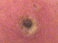
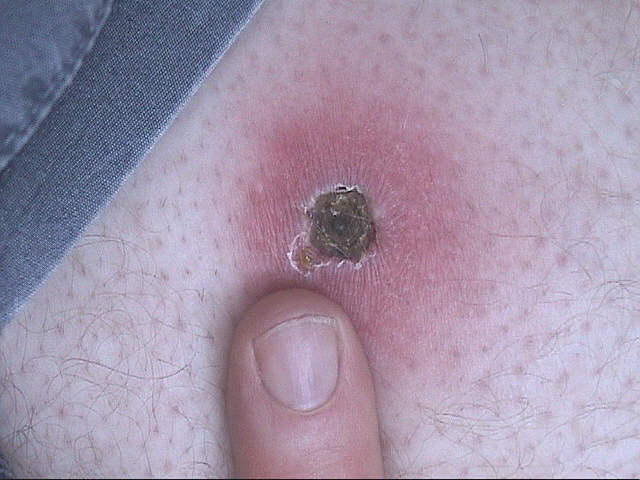
 Eschar Formation
 -- This photo at left was marked "Day 3 -- Pus & Redness."
The initial application of
Cansema Salve
to skin that contains cancer cells produces a rubifacient (reddening) effect,
some edema, and quite often a pain response - usually mild. (The degree of
all three of these type responses will depend greatly on how much cancer
activity there is on the target site, how close to the surface of the skin,
and other factors, including immunological conditions, that are unique
to the user/patient).
These response characteristics are not
necessarily unique to
Cansema. In fact, most products that
are in the same class of
escharotic preparation
will cause similar response(s).
(Note --- novices in this area would do well to read our
FAQ
section on Cansema & escharotics for a quick briefing on
the general subject).
The photo at the right, above, is marked
"Day 5" and shows an eschar formation that is well on the way to decavitation.
Notice that both the eschar and the surrounding healthy tissue appear "dried out,"
with the edematous appearance now gone. The rubifacient look is still there,
so there is still irritation and a high leukocyte count in the surrounding tissue.
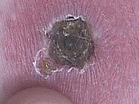
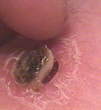
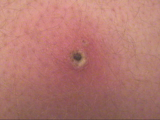
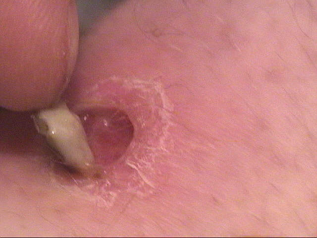
 Eschar Formation, Late Stage
 --
Here we have an excellent photo showing the eschar in its late stage.
It has dried up to the point where it has begin to pull away from the
surrounding, healthy tissue, from which its surface is elevated.
Note that both the edema and rubifacience have died down
considerably. The immune system has done its job -- so,
from the body's point of view, the eschar ejection is almost
mechanical. The underlying dermal layers will continue to
grow in and push out the necrosed mass, with the top layer
becoming the new
stratum
corneum.
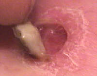
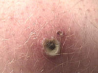
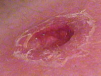
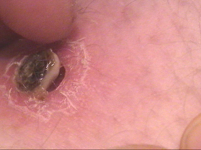
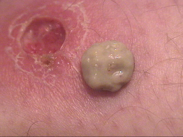
 Eschar Removal
 -- More excellent photos showing eschar
removal -- which then initiates the decavitation stage.
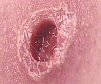
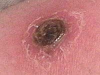
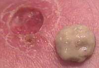
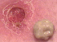
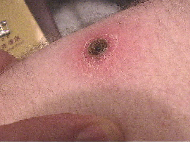
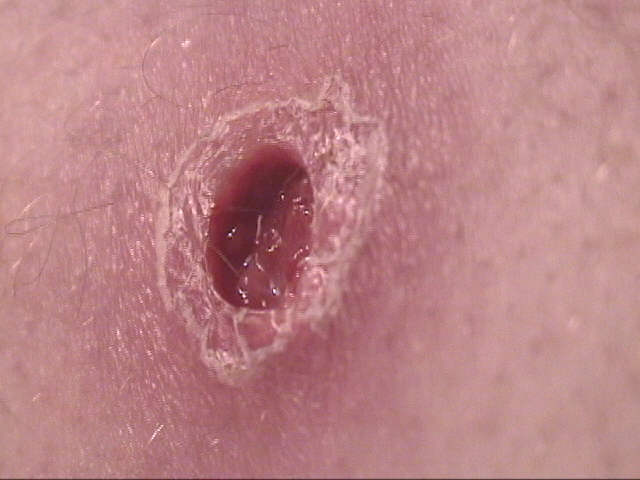
 Decavitation
 -- Photos such as this one clearly demonstrate the
discriminating characteristics of Cansema. The eschar encorporated
the entirety of the cancer (or the entire localized cancer growth --
we have no way of knowing if there is cancer elsewhere in the body),
leaving the surrounding, healthy tissue only mildly irritated.
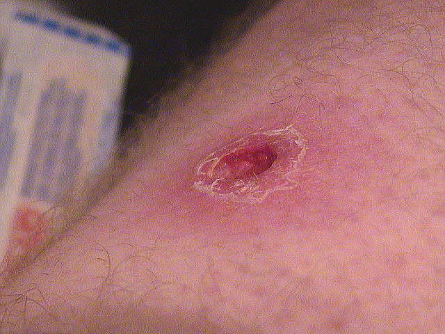
Heal Over -- Although there were no "heal over" photographs
with this case, the progression to this stage has been photographed
elsewhere. The completion of this phase, with the regrowth of
the epidermal tissue, and fading of hyperpigmentation. Most
cases involve little or no scar tissue, but some cases, particularly
where the growth was deep and/or successive Cansema applications
were required or performed, may involve some scar tissue.

 Eschar Formation -- This photo at left was marked "Day 3 -- Pus & Redness."
The initial application of
Cansema Salve
to skin that contains cancer cells produces a rubifacient (reddening) effect,
some edema, and quite often a pain response - usually mild. (The degree of
all three of these type responses will depend greatly on how much cancer
activity there is on the target site, how close to the surface of the skin,
and other factors, including immunological conditions, that are unique
to the user/patient).
Eschar Formation -- This photo at left was marked "Day 3 -- Pus & Redness."
The initial application of
Cansema Salve
to skin that contains cancer cells produces a rubifacient (reddening) effect,
some edema, and quite often a pain response - usually mild. (The degree of
all three of these type responses will depend greatly on how much cancer
activity there is on the target site, how close to the surface of the skin,
and other factors, including immunological conditions, that are unique
to the user/patient).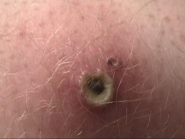
 Eschar Removal -- More excellent photos showing eschar
removal -- which then initiates the decavitation stage.
Eschar Removal -- More excellent photos showing eschar
removal -- which then initiates the decavitation stage.
 Decavitation -- Photos such as this one clearly demonstrate the
discriminating characteristics of Cansema. The eschar encorporated
the entirety of the cancer (or the entire localized cancer growth --
we have no way of knowing if there is cancer elsewhere in the body),
leaving the surrounding, healthy tissue only mildly irritated.
Decavitation -- Photos such as this one clearly demonstrate the
discriminating characteristics of Cansema. The eschar encorporated
the entirety of the cancer (or the entire localized cancer growth --
we have no way of knowing if there is cancer elsewhere in the body),
leaving the surrounding, healthy tissue only mildly irritated.